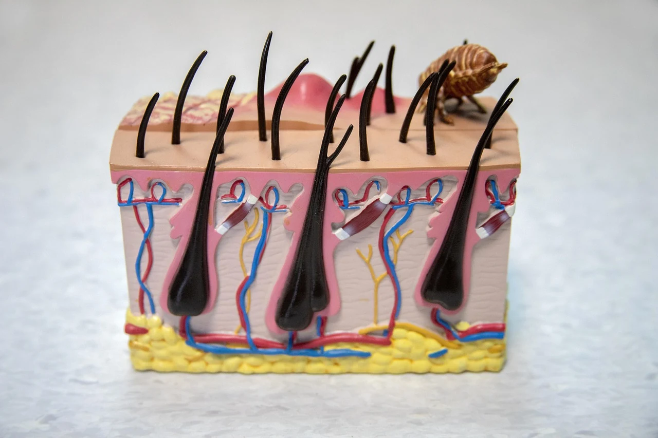

An atheroma is a rounded growth under the skin that occurs due to blockage of a sebaceous gland duct. In English-language reference books it is described as an epidermoid cyst, and the term "sebaceous cyst" is considered inaccurate. Below is what this growth is and when a surgeon is needed.
Atheroma: What It Is and How It Differs from Other Cysts
An atheroma, or sebaceous gland cyst, is a tumor-like growth that occurs when a sebaceous gland duct becomes blocked. It is a benign cyst arising from the infundibulum or upper part of a hair follicle, filled with keratin and lipid debris.
The familiar sebaceous cyst is more accurately called an epidermoid cyst: its cavity is filled with desquamated skin cells. A true sebaceous gland cyst — a steatocystoma — contains yellowish oily material.
Why Sebaceous Gland Blockage Occurs
The main mechanism is blockage of a hair follicle along with its sebaceous gland. In areas without hair (buttocks, palms, soles), a cyst forms when epidermal cells are traumatically introduced into the dermis.
The cause is generally unknown; in some people, relatives had the same growths. They occur more often in adults than in children, predominantly in young and middle age, and in men twice as often as in women. Multiple cysts have been described in acromegaly and hereditary syndromes, such as Gardner syndrome. Secondary atheromas occur in people with oily seborrhea and severe acne.
What an Atheroma Looks Like and Its Symptoms
The typical picture is a firm nodule of flesh-colored or yellowish color, usually 1–3 cm in diameter, fixed to the skin but movable relative to deeper tissues. A punctate pore with cheesy discharge is often visible in the center. The nodule is usually not painful and grows slowly, and once it reaches a certain size, it stops enlarging.
Common locations:
- chest, shoulders, and back;
- face, especially below the mouth, and scalp;
- neck and retroauricular fold;
- skin of the genital organs, including the scrotum and vulva.
Secondary atheromas are firmer, more painful, with a bluish tint and a size ranging from a pea to a hazelnut; more often on the cheeks, neck, and chest.
Whether an Atheroma Is Dangerous: Inflammation and Complications
It is usually a benign growth. The main problem is inflammation: when the contents rupture into the dermis, swelling, redness, and tenderness appear. The cause is trauma or bacterial infection, most often Staphylococcus aureus and group A Streptococcus. An inflamed cyst suppurates and forms a painful abscess with fever and fluctuation; sometimes the growth ruptures with purulent discharge. After incomplete removal of the capsule, the cyst recurs. Extremely rarely, squamous cell skin cancer develops inside such a cyst.
How a Doctor Makes the Diagnosis and Distinguishes an Atheroma from Other Growths
The diagnosis is made on examination: the shape, central pore, and location are recognized without additional tests. A biopsy is not needed in typical cases. If infection is suspected, a skin culture is taken. Ultrasound is used for initial evaluation of a soft-tissue growth; for a typical atheroma it is usually not required.
The differential series includes lipoma (a movable node 2–10 cm in size of soft consistency), trichilemmal cyst (a firm node 0.5–5 cm without a central pore, more often on the scalp), pseudocyst in acne, dermoid cyst in children, and epidermal cyst associated with human papillomavirus. Distinguishing an atheroma from other benign skin growths can sometimes be difficult.
When to See a Surgeon
The reason for consultation is any new growth on or under the skin: the doctor rules out a malignant process. An unscheduled visit is needed if the cyst has become red, painful, or swelling or fever has appeared — these are signs of inflammation or suppuration. A surgeon or dermatologist assesses the size, mobility, skin condition, and signs of inflammation, after which they determine whether observation or removal is needed.
An atheroma by itself is not a reason to call an ambulance; 103/112 — if the condition sharply worsens and there is a threat to life (for example, severe weakness, loss of consciousness, difficulty breathing against the background of inflammation).
This material is for reference: it describes signs and the order of actions, but does not replace examination by a doctor.
Sources
Update history
Reader reviews
No reviews yet — tell us whether this article was helpful.
Leave a review
This content is for reference and does not replace a doctor's consultation. Medical disclaimer